Tendon Diagram / Biology And Anatomy Of Flexor Tendon Youtube. To bend the elbow and to turn the palm of the hand towards the sky. One of the most important tendons in terms of mobility of the leg is the achilles tendon. Learn about the anatomy and physiology of tendons. Ligaments connect one bone to another, while tendons connect muscle to bone. The patellar tendon holds the patella with other two bones, similarly iliotibial band helps in supporting tibia and fibula.
The achilles tendon is the largest. (b) a photomicrograph of the tissue and (c) a labeled diagram. Hand a hand is a prehensile multi fingered appendage located at the end of the forearm or forelimb of primates such as humans chimpanzees monkeys and lemurs human anatomy for the artist the dorsal hand the dorsal the easiest tendons to identify in the dorsal hand are those of the extensor digitorum muscle its name means extensor of the digits which is One of the most important tendons in terms of mobility of the leg is the achilles tendon. Tendon, tissue that attaches a muscle to other body parts, usually bones.
Ankle Anatomy Eorthopod Com from eorthopod.com Tendons are tough, flexible, fibrous bands of tissue that connect muscles to bones. The foot diagram has a complex structure made up of bones, ligaments, muscles, and tendons.understanding the structure of the foot is best done by looking at a foot diagram where the anatomy has been labeled. A tendon, also known as a sinew, is a fibrous tissue that helps to facilitate this movement. (b) a photomicrograph of the tissue and (c) a labeled diagram. Thirty percent of all people will have a tendon injury, and the risk is higher in women, she says. The main difference between tendons and ligaments is that they connect different parts of the anatomy. The achilles tendon is the largest. Learn about the anatomy and physiology of tendons.
Tendon definition, a cord or band of dense, tough, inelastic, white, fibrous tissue, serving to connect a muscle with a bone or part;
The tendons have 2 functions: 1 tendons join muscles to their corresponding bones. One tendons inserts onto the forearm bone, the radius, and the second spreads out to join the fascia along the upper part of the forearm. Tendons are viscoelastic structures, which means they exhibit both elastic and viscous behaviour. The anterior cruciate ligament prevents the femur from sliding backward on the tibia (or the tibia sliding forward on the femur). Tendons that make this possible include: This important tendon in the back of the calf and ankle stores the elastic energy needed for running, jumping, and other physical activity. Don't forget to share this picture with others via. Related posts of diagram of shoulder muscles and tendons frog muscle anatomy. A tendon, also known as a sinew, is a fibrous tissue that helps to facilitate this movement. Tendons in the knee play a very important role in holding the knee and the muscles together. Ligaments join the knee bones and provide stability to the knee: Tendon, tissue that attaches a muscle to other body parts, usually bones.
The thoracic diaphragm, or simply the diaphragm (ancient greek: Hand a hand is a prehensile multi fingered appendage located at the end of the forearm or forelimb of primates such as humans chimpanzees monkeys and lemurs human anatomy for the artist the dorsal hand the dorsal the easiest tendons to identify in the dorsal hand are those of the extensor digitorum muscle its name means extensor of the digits which is This important tendon in the back of the calf and ankle stores the elastic energy needed for running, jumping, and other physical activity. Broadly considered, human muscle—like the muscles of all vertebrates—is often divided into striated muscle, smooth muscle, and cardiac muscle. Allows the action of raising the foot.
Leg Muscle And Tendon Diagram Google Search Leg Muscles Anatomy Muscle Anatomy Body Anatomy from i.pinimg.com Allows the action of raising the foot. Tendons that make this possible include: Frog muscle anatomy 12 photos of the frog muscle anatomy frog leg muscle anatomy. The tendon that attaches the biceps muscle to the forearm bones (radius and ulna) is called the distal biceps tendon. The patella is a sesamoid bone that lies within the quadriceps tendon. (b) a photomicrograph of the tissue and (c) a labeled diagram. 'partition'), is a sheet of internal skeletal muscle in humans and other mammals that extends across the bottom of the thoracic cavity.the diaphragm separates the thoracic cavity, containing the heart and lungs, from the abdominal cavity and performs an important function in. Tendons are sometimes confused with ligaments.
To bend the elbow and to turn the palm of the hand towards the sky.
Ligaments and tendons serve similar purposes, but in different ways. One tendons inserts onto the forearm bone, the radius, and the second spreads out to join the fascia along the upper part of the forearm. Tendons are found throughout the body, from the head and neck all the way down to the feet. Ligaments are tough, flexible bands of connecting tissue that join bones to other bones. To bend the elbow and to turn the palm of the hand towards the sky. Attaches the calf muscles to the calcaneus, most important muscles for running, jumping, walking etc. Related posts of foot tendons and ligaments diagram gastrocnemius muscle anatomy. The thoracic diaphragm, or simply the diaphragm (ancient greek: Arm tendon diagram the difference between a normal switch and a three way switch is 1 more arm tendon diagram because the travellers or messenger terminals are usually interconnected, the. Tendons in the knee play a very important role in holding the knee and the muscles together. Human muscle system, the muscles of the human body that work the skeletal system, that are under voluntary control, and that are concerned with movement, posture, and balance. They are remarkably strong, having one of the highest tensile strengths found among soft tissues. Ligaments connect one bone to another, while tendons connect muscle to bone.
When the biceps contracts, it pulls the forearm up and rotates it outward. 'partition'), is a sheet of internal skeletal muscle in humans and other mammals that extends across the bottom of the thoracic cavity.the diaphragm separates the thoracic cavity, containing the heart and lungs, from the abdominal cavity and performs an important function in. When stretched, tendons exhibit typical soft tissue behavior. Tendons are prone to injuries caused by overuse. Two of the main ligaments in the back are the anterior longitudinal ligament and the posterior longitudinal.
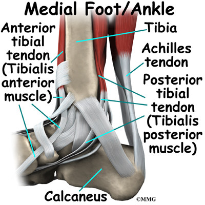
Tendon Rupture Anatomical Example Vector Illustration Diagram Vectormine from selzimg.s3.amazonaws.com The patellar tendon connects the apex of the patella to the tibial tuberosity, and improves the way the quadriceps muscle pulls on the tibia. Human muscle system, the muscles of the human body that work the skeletal system, that are under voluntary control, and that are concerned with movement, posture, and balance. Ligaments join the knee bones and provide stability to the knee: Tendons attach or support the joints between muscles and bones, while ligaments support the joints between bones. In the leg muscles diagram above, there are many muscles that make up your legs and support it to move. Allows the foot to be turned inward and also supports the arch of the foot. Ligaments and tendons serve similar purposes, but in different ways. One tendons inserts onto the forearm bone, the radius, and the second spreads out to join the fascia along the upper part of the forearm.
The patellar tendon connects the apex of the patella to the tibial tuberosity, and improves the way the quadriceps muscle pulls on the tibia.
The anterior cruciate ligament prevents the femur from sliding backward on the tibia (or the tibia sliding forward on the femur). Ligaments are tough, flexible bands of connecting tissue that join bones to other bones. The thoracic diaphragm, or simply the diaphragm (ancient greek: 'partition'), is a sheet of internal skeletal muscle in humans and other mammals that extends across the bottom of the thoracic cavity.the diaphragm separates the thoracic cavity, containing the heart and lungs, from the abdominal cavity and performs an important function in. The main difference between tendons and ligaments is that they connect different parts of the anatomy. Two of the main ligaments in the back are the anterior longitudinal ligament and the posterior longitudinal. Tendon definition, a cord or band of dense, tough, inelastic, white, fibrous tissue, serving to connect a muscle with a bone or part; Tendons, located at each end of a muscle, attach muscle to bone. 2 ligaments (trapezoid& conoid ligaments) attach the clavicle coracoid process of scapula these tiny ligaments (w/ acominoclavicular joint) keep scapula attached to clavicle. When the biceps contracts, it pulls the forearm up and rotates it outward. The muscle belly then crosses the entire upper arm and separates into two tendons. When tendons become inflamed, irritated or suffer microscopic tears, the condition is called tendonitis. Attaches the calf muscles to the calcaneus, most important muscles for running, jumping, walking etc.
Share :
Post a Comment
for "Tendon Diagram / Biology And Anatomy Of Flexor Tendon Youtube"


Post a Comment for "Tendon Diagram / Biology And Anatomy Of Flexor Tendon Youtube"